Abstract
Most people are not aware that the soil around them is a battle scene. The combatants are very small—bacteria on one side and bacteriophage on the other. The bacteriophage (or phage for short) try to pierce the outer coats of the bacteria and inject them with phage DNA. If successful, the DNA will take over the inner machinery of the bacterial cells and force them to make many copies of the phage. After the copies are made, the bacterial cells break apart, releasing new phage that start the hunt all over again. The fact that phage can kill bacteria has led to the suggestion that phage could be used to fight human bacterial infections. In this science project, you will experiment with phage infection of E. coli bacteria. But you won't have to dig around in the dirt; the experiments will be done in a lab and the bacteria and phage will be grown in petri dishes.Summary
David B. Whyte, PhD, Science Buddies
This science project was based on this Science Buddies Clever Scientist Award winning project:
- Obright, J. (2010). A Comparison of the Effects of Streptomycin and Coliphage Lambda on Escherichia coli. Displayed at the Minnesota State and Science Engineering Fair, March 2010.

Objective
Infect growing E. coli B cultures with bacteriophage T4r and demonstrate that the number of plaques formed can vary from a few to innumerable. Demonstrate that phage can effectively kill E. coli and could possibly be used to fight E. coli infections.
Introduction
Bacteria are microscopic, single-celled organisms found in air, water, soil, and food. They live on plants, insects, animals, pets, and even in the human digestive system and upper respiratory tract. There are thousands of kinds of bacteria. Most are not associated with diseases in humans, but some, such as Escherichia coli O157:H7 (E. coli O157:H7) can cause serious illnesses. E. coli O157:H7 has caused several nationally prominent outbreaks of food poisoning. An estimated 73,000 cases are reported in the United States every year. The "bug" is transmitted through contaminated food, such as hamburger meat and unwashed fruits and vegetables.
Antibiotics are the mainstay of bacterial treatment. The goal of these drugs is to kill invading bacteria without harming the host. When antibiotics were discovered in the 1940s, they were very effective in bacterial infection treatment. Over time, however, many antibiotics have lost effectiveness against common bacterial infections because of increasing drug resistance. Bacteria might be naturally resistant to different classes of antibiotics or might acquire resistance from other bacteria through exchange of resistant genes. Indiscriminate, inappropriate, and prolonged use of antibiotics have created antibiotic-resistant bacteria. Antibiotic-resistant strains, which have emerged in hospitals, long-term care facilities, and communities worldwide, pose a real medical challenge.
The goal of this science project is to experiment with the use of bacteriophage to kill E. coli bacteria. Bacteriophages (or phages, for short) are small viruses that infect bacteria. Because they can kill the bacteria that they infect, it is possible that phages could be used to combat antibiotic-resistant strains of bacteria. The phage you will use is called T4r, a member of the T4 phages. T4 phages are lytic phages, meaning that they lyse (break open) the bacterial cells that are infected. See Figure 1 for an image of a phage, and the video below the figure for an animation of how they break open a cell. Their structure looks like something out of a science fiction novel!
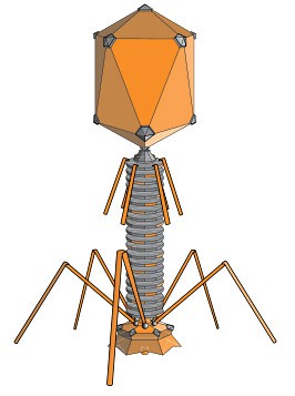 Image Credit: Wikipedia / Creative commons
Figure 1. Structure of a T4 phage. The phage attaches to the surface of a bacterial cell and injects DNA into the interior of the cell. T4 infects E. coli. Other phages infect other kinds of bacteria. The phage DNA "hijacks" the bacteria's cellular machinery, forcing it to produce many new copies of the phage. When the new phage particles have been assembled, they burst out of the bacteria and start the cycle over again. (Wikipedia, 2010.)
To grow the phage, you will mix phage with a liquid suspension of E. coli cells. For the sake of safety, in this science project you will use the safe laboratory strain E. coli B in place of the pathogen E. coli O157:H7. The mixture of E. coli and phage will be spread out onto the surface of an agar plate. The E.coli will grow to form a visible layer on the agar. This layer is called a bacterial lawn. If a phage has attached to an E.coli cell and has successfully reproduced, it will form a relatively clear circle in the lawn. The circle forms because the phage have broken open the E. coli cells in this area. The clear area on the bacterial lawn formed by the phage is called a plaque. A single plaque can contain many thousands of new phage particles, all derived from a single phage particle. By counting the number of plaques formed, and knowing the dilution used, you can calculate the number of plaque-forming units (PFUs) per milliliter in the phage stock. For example, if 0.1 mL of a 1:1,000 dilution of a phage stock produces 100 plaques, the stock has 100,000 PFUs per 0.1 mL. In order to titer a phage stock—that is, to measure the number of PFUs per unit volume—several dilutions of a phage stock will be mixed with E. coli and plated onto agar. Several dilutions will be used so that at least one plate will have a good number of plaques to count. What is a good number? If there are less than 10 per plate, the titer will be inaccurate; one plaque more or less changes the titer by 10 percent! If there are too many plaques, they will merge, and the whole lawn of E. coli will be lysed. Since the focus of this project is the use of phage to effectively kill all of the bacteria, the goal will be to determine the minimum "dose" of phage that should be used to kill virtually all of the E. coli on the plate. In order to find the right dose, you will have to obtain an accurate titer by plating phage dilutions and counting plaques. Let's get started!
Terms and Concepts
- Bacteria
- Escherichia coli O157:H7 (E. coli O157:H7)
- E. coli B
- Antibiotics
- Drug-resistance
- Antibiotic-resistant bacteria
- Bacteriophage
- Phage
- T4 phage
- Lytic infection
- Agar plate
- Lawn of bacteria
- Plaque
- Plaque-forming units (PFUs)
- Titer
Questions
- What is a bacterial strain? How are bacterial strains E. coli O157:H7 and E. coli B similar? How are they different? Which one are you using for your experiment and why?
- Where are phage found in the environment?
- Based on your research, does a particular kind of phage tend to infect just one kind of bacteria, or more than one?
- Based on your research, what phages form lytic infections of E. coli?
- Based on your research, are phages now being used in medical procedures to fight bacterial infections?
- What are the advantages and disadvantages of using phages to fight infections, compared to antibiotics?
- What does it mean to "titer a phage stock"?
- What does the word coliphage mean?
- What is the difference between a phage and a plaque-forming unit? Hint: Not all phage form plaques.
Bibliography
- Mayer, G. (2010). Bacteriophage. Retrieved April 27, 2010.
- Cells Alive. (2006). Oh Goodness! My E. coli has a Virus!. Retrieved April 27, 2010.
Materials and Equipment
Note: It will be a very helpful if you have access to a lab with pipets, petri dishes, cotton swabs, and other common supplies. Talk to your teachers at school to obtain access.
-
Coliphage culture and titer determination study kit; available from Ward's Science Notes: 1) This kit has enough supplies to go through the procedure one time. Purchase additional materials for repeat trials, which will help ensure your results are accurate and repeatable. This is an advanced procedure, so the details of how to repeat the procedure are not supplied. 2) This kit contains the strain E. coli B which is not a human pathogen and is safe to use in a classroom laboratory. Standard bacterial safety, detailed below, should still be followed.
- Culture of E. coli B
- Culture of T4r phage
- Tryptic soy agar (125 mL)
- 2-mL tubes of soft agar (6)
- 9-mL dilution tubes (12)
- 1- x 0.01-mL sterile pipets (12)
- Sterile petri dishes (6)
- Sterile swab
- Pipet bulb
-
Extra supplies to repeat the procedure; available from Ward's Science at www.wardsci.com:
- Tryptic soy agar
- Soft soy agar
- Tryptic soy broth
- Petri dishes
- Permanent marker
- Bacterial incubator, 37°C
- Microwave
- Thermometer
- Water bath, 42°C
- Beaker, 1-L; should be made of a material that can hold boiling water
- Lab notebook
Disclaimer: Science Buddies participates in affiliate programs with Home Science Tools, Amazon.com, Carolina Biological, and Jameco Electronics. Proceeds from the affiliate programs help support Science Buddies, a 501(c)(3) public charity, and keep our resources free for everyone. Our top priority is student learning. If you have any comments (positive or negative) related to purchases you've made for science projects from recommendations on our site, please let us know. Write to us at scibuddy@sciencebuddies.org.
Experimental Procedure
For health and safety reasons, science fairs regulate what kinds of biological materials can be used in science fair projects. You should check with your science fair's Scientific Review Committee before starting this experiment to make sure your science fair project complies with all local rules. Many science fairs follow Regeneron International Science and Engineering Fair (ISEF) regulations. For more information, visit these Science Buddies pages: Project Involving Potentially Hazardous Biological Agents and Scientific Review Committee. You can also visit the webpage ISEF Rules & Guidelines directly.
- Use a sterile cotton swab to transfer a small quantity of the bacteria from the slant culture to a broth tube (9-mL dilution tube).
- Place the tube in the 37°C incubator overnight, with the top loosened.
- Loosen the cap on the 125-mL agar bottle and heat the bottle in a microwave until the agar is melted.
- Pour the molten agar into six petri dishes.
- After the plates have hardened, turn them upside-down and leave them at room temperature overnight.
- Label the plates Control and 10-6 through 10-10.
30 Minutes Before the Experiment: Make Your Phage Dilutions.
- Add water to the beaker until it is about half full.
- Heat the water in the microwave until it is boiling.
- Place the six soft tryptic soy agar tubes into the beaker of hot water and loosen the caps half a turn.
- Wait for the agar to melt. Microwave the water to boiling again, if needed.
-
When the agar has melted, transfer the tubes to a 42°C water bath.
- At this temperature, the agar will stay liquid and the E. coli can be added without being harmed by high temperature.
- Label the soft agar tubes Control and 10-6 through 10-10.
- Label 11 of the broth tubes Control and 10-1 through 10-10. These tubes have 9.0 mL of broth in them.
- Pipet 1.0 mL of the phage broth up and down 8–10 times to mix it.
- Transfer 1.0 mL of the phage broth into the broth tube labeled 10-1. Note: Make sure you understand how this dilution, and all subsequent ones, are calculated; sit down and work it out on a piece of paper. If you're still confused consult a math or science teacher.
- Use a fresh pipet to transfer 1.0 mL from the broth tube labeled 10-1 to the broth tube labeled 10-2.
- Continue in this manner until you have pipeted from the 10-4 to the 10-5 broth tube.
-
With a fresh pipet, withdraw 1.1 mL phage and broth from the 10-5 broth tube.
-
Add 0.1 mL to the 10-6 soft agar tube.
- This is equivalent to adding 1.0 mL of 10-6 phage. The lower volume helps the agar stay firm after it cools.
- Soft agar has a lower concentration of agar than is used to make the agar petri dishes. This keeps the E. coli and phage from moving freely, but is soft enough to allow for bacterial growth.
- Add the remaining 1.0 mL of phage and broth to the 10-6 broth tube, mixing as above.
-
Add 0.1 mL to the 10-6 soft agar tube.
- With a fresh pipet, withdraw 1.1 mL from the 10-6 broth tube and add 0.1 mL to the 10-7 soft agar tube and 1.0 mL to the 10-7 broth dilution tube.
- Continue in this manner until you reach the 10-10 soft agar and broth dilution tubes.
- Add 0.25 mL of E. coli broth culture to each soft agar tube, using a clean pipet.
- After the E. coli has been added, pour the contents of each tube onto the agar surface of the corresponding plate.
- Immediately after pouring each tube, swirl the plate to distribute the soft agar over the surface.
- Pipet 0.1 mL of the blank (no phage) tryptic soy broth into the control soft agar tube containing E. coli and pour this onto the blank control petri dish.
- After the overlays have hardened (1–2 hours), place the plates in an inverted position in a 37°C incubator for 12–24 hours.
- Plaques should form 10–24 hours after plating.
Counting the Plaques
- Make a data table, with the dilutions you used in one of the columns.
- Count the number of plaques for each dilution. If there are too many to count, just put "TMTC" in the table.
-
Determine the titer.
- Use plates that have between 30 and 300 plaques for your calculations.
- For example, if the plate labeled 10-7 has 100 plaques, the titer of the stock suspension is 100 x10 +7 PFU per mL, or 109 PFU per mL.
- Perform the entire procedure two more times with fresh materials. This demonstrates that your results can be repeated.
Analyzing Your Data
- How many PFUs did you have to add to lyse the entire lawn of E. coli?
- Now that you have worked with phage, what do you think are the pros and cons of using phage to treat bacterial infections?
Bacterial Safety
Bacteria are all around us in our daily lives and the vast majority of them are not harmful. However, for maximum safety, all bacterial cultures should always be treated as potential hazards. This means that proper handling, cleanup, and disposal are necessary. Below are a few important safety reminders.
- Keep your nose and mouth away from tubes, pipettes, or other tools that come in contact with bacterial cultures, in order to avoid ingesting or inhaling any bacteria.
- Make sure to wash your hands thoroughly after handling bacteria.
- Proper Disposal of Bacterial Cultures
- Bacterial cultures, plates, and disposables that are used to manipulate the bacteria should be soaked in a 10% bleach solution (1 part bleach to 9 parts water) for 1–2 hours.
- Use caution when handling the bleach, as it can ruin your clothes if spilled, and any disinfectant can be harmful if splashed in your eyes.
- After bleach treatment is completed, these items can be placed in your normal household garbage.
- Cleaning Your Work Area
- At the end of your experiment, use a disinfectant, such as 70% ethanol, a 10% bleach solution, or a commercial antibacterial kitchen/bath cleaning solution, to thoroughly clean any surfaces you have used.
- Be aware of the possible hazards of disinfectants and use them carefully.
Ask an Expert
Global Connections
The United Nations Sustainable Development Goals (UNSDGs) are a blueprint to achieve a better and more sustainable future for all.
Variations
- How does temperature affect the growth of plaques?
- How does the time of incubation affect the growth of plaques?
- Try other phage types.
- Add E. coli and phage dilutions to raw hamburger. Does the phage kill the bacteria in this situation? Hint: Try counting E. coli colonies in extracts from the "contaminated" meat.
Careers
If you like this project, you might enjoy exploring these related careers:
Related Links
- Science Fair Project Guide
- Other Ideas Like This
- Microbiology Project Ideas
- My Favorites
- Microorganisms Safety Guide
- Microbiology Techniques and Troubleshooting
- Scientific Review Committee


