/-/https/www.sciencebuddies.org/Files/21464/5/sb-logo.png)
/-/https/www.sciencebuddies.org/Files/21464/5/sb-logo.png)
Cell Organization in the Human Body
Summary
/-/https/www.sciencebuddies.org/Files/17088/6/cell-organization-lung-model.jpg)
Overview
The human body is complex! In this lesson, it is broken down into smaller, manageable parts. Students will build a lung model and discover how different parts of the respiratory system work together to make breathing possible. Next, they will discuss how this system works together with other systems in the body so oxygen can reach every cell of the body. While students perform these explorations, they will realize that multicellular organisms consist of systems, which are a collection of organs and tissues, and all of these are made of cells.
Remote Learning: This lesson plan can be adapted to work remotely. The Engage section of the lesson can be done over a video call. Then, students can make the model individually and independently using the Make a More Advanced Lung Model video as a guide. Allow the students to make a model with only one lung instead of two as joining two straws can be tricky. A set of materials can be prepared in advance, or, if students gather the materials themselves, ask for adult supervision while cutting the hard plastic bottle. Students can use the Student Worksheet to complete their exploration independently and can then share their observations and thoughts with each other, virtually. You can also use the Simple Lung Model video to help students understand the breathing mechanism shown in the model. End the lesson with a discussion over a video call during the Reflect section.
Learning Objectives
- Understand the different levels of organization (cell, tissue, organ, system) in an organism.
- List examples of cells being organized into tissues, organs, and systems in the human body (e.g. lung cell, lung tissue, the lung, respiratory system).
- Understand how the human respiratory system works, and how it interacts with the human circulatory system.
NGSS Alignment
This lesson helps students prepare for these Next Generation Science Standards Performance Expectations:- MS-LS1-3. Use argument supported by evidence for how the body is a system of interacting subsystems composed of groups of cells.
|
Science & Engineering Practices
Engaging in Argument from Evidence.
Use an oral and written argument supported by evidence to support or refute an explanation or a model for a phenomenon.
Developing and Using Models. Develop and/or use a model to predict and/or describe phenomena. Develop a model to describe unobservable mechanisms. |
Disciplinary Core Ideas
LS1.A: Structure and Function.
In multicellular organisms, the body is a system of multiple interacting subsystems. These subsystems are groups of cells that work together to form tissues and organs that are specialized for particular body functions.
|
Crosscutting Concepts
Systems and System Models.
Systems may interact with other systems; they may have sub-systems and be a part of larger complex systems.
A system can be described in terms of its components and their interactions. Structure and Function. Complex and microscopic structures and systems can be visualized, modeled, and used to describe how their function depends on the shapes, composition, and relationships among its parts, therefore complex natural structures/systems can be analyzed to determine how they function. |
Materials
/-/https/www.sciencebuddies.org/Files/17089/6/Advanced-lung-model-materials.jpg)
Per group of 2–4 students:
- Disposable, empty transparent bottle (10–16 fluid oz) with lid, made of hard plastic (such as a sports drink bottle)
- Three balloons (8-inch or 6-inch balloons work well)
- Paper or plastic drinking straw
- Tape
- Scissors
Teacher or another adult:
- Utility knife
- A few extra balloons in case one pops
- A few extra straws
Background Information for Teachers
This section contains a quick review for teachers of the science and concepts covered in this lesson.All living organisms consist of cells, but most multicellular organisms are not simply a group of cells. Several organizational structures can be found within these organisms. In increasing level of complexity, cells are grouped into tissues, organs, and systems before reaching the level of the organism. This lesson will use the human body to explain this organization.
Organs and other parts of the body that work together to perform a major function in a living organism are called systems. We can identify ten systems in the human body, each with its own distinct task: the circulatory or cardiovascular system pumps blood through the body, the respiratory system allows the exchange of gases, the digestive system breaks down food for energy production, the endocrine system controls the hormones in our bodies, the reproductive system allows reproduction, the muscular system helps with the movement of the body, the nervous system controls the senses, the urinary system allows the removal of waste, the skeletal system gives the body its structural support, and the integumentary system creates protection through the skin, hair, and nails. Each of these systems consists of several parts. The respiratory system, for example, consists of the nose with the sinuses and nasal cavity, the pharynx, the larynx or voice box, the trachea or windpipe, the bronchi and bronchia, the lungs with the alveoli, the pulmonary capillaries, and the diaphragm (Figure 1). Note that the mouth and some muscles other than the diaphragm also aid breathing, but are listed under the digestive system and the muscular system, respectively. Each part of the respiratory system fulfills a specific task, and it is made of tissue that helps fulfill this task. The diaphragm is a muscle and made of muscle cells that together form muscle tissue; similarly, the lungs are made up of lung cells and make up lung tissue, and the windpipe is made of cartilage, yet another type of tissue. Yet, all these parts work together to support breathing.
/-/https/www.sciencebuddies.org/Files/17092/7/Respiratorysystem.png)
Figure 1. Visualization of the respiratory system with all its parts.
Each system also needs the help of other systems to fulfill its task inside the body. The respiratory system does not work in solitude, it relies on the circulatory system to transport oxygen to the cells and to transport carbon dioxide back to the lungs.
Because the human body is a complex multilevel organization, minor deviations can have seemingly unrelated consequences. In this lesson, students will explore this by looking into iron deficiency anemia. Iron is needed to create hemoglobin, the oxygen-carrying protein that give red blood cells their red color. Insufficient iron leads to insufficient hemoglobin and an inadequate way of delivering oxygen to the cells around the body. It can show as fatigue, or even shortness of breath.
In this lesson, students will create a model of the respiratory system to discover how it works. This allows students to see how tissues are groups of similar cells that are specialized in a particular function, and how organs are groups of specialized tissues that are gathered in a particular area in the body and perform a specific function. Students will see how the respiratory system works, and how it needs the help of other systems. In addition, students will learn how, on a cellular or even molecular level, oxygen is carried around in the body. With all this information, students can finally start to comprehend why iron-deficient anemic patients may suffer from shortness of breath.
As students dive into a particular system and zoom out to see how the systems work together, they will hopefully gain some respect for the complexity, beauty, and efficiency of their bodies.
Additional Background Links
Prep Work (20 minutes)
- Prepare a bottle, one for each group of students.
- Empty and clean a transparent, disposable bottle made of hard plastic.
- Cut the bottom part of the bottle off.
- Make a hole in the middle the bottle cap, just large enough for a straw.
- Keep the cut-off bottle and the cap together.
- Watch the instructional video below. It explains how to make a simple lung model and how it models human breathing. This lesson will result in the more-advanced model, shown at timestamp 2:02 in this video. This video clearly explains how these models mimic the breathing process.
Create AssignmentEmbed Link
 Google Classroom
 Microsoft Teams
 Canvas
 Schoology
 Other
Other - Optional: Make a simple or more-advanced lung model yourself to show the students. The video on how to make the more-advanced lung model is included in the Explore section.
/-/https/i.ytimg.com/vi/WJ06mrNliC0/maxresdefault.jpg)
/-/https/www.sciencebuddies.org/Files/20511/4/google-classroom.png)
/-/https/www.sciencebuddies.org/Files/20512/4/ms-teams.png)
/-/https/www.sciencebuddies.org/Files/20469/4/Canvas_Bug_Color_RGB.png)
/-/https/www.sciencebuddies.org/Files/20470/5/Schoology-Logo.png)
/-/https/www.sciencebuddies.org/Files/20471/4/lms.png)
/-/https/www.sciencebuddies.org/Files/20469/4/Canvas_Bug_Color_RGB.png)
/-/https/www.sciencebuddies.org/Files/20470/5/Schoology-Logo.png)
/-/https/www.sciencebuddies.org/Files/20471/4/lms.png)
Teacher Tool Box
Engage (15 minutes)
- Introduce anemia as a phenomenon.
- Ask students if they know what iron deficiency anemia is. If they do not know, explain that it is a condition where a deficiency in iron leads to anemia, or a reduced amount of red blood cells or a reduced amount of hemoglobin in the red blood cells.
- Show the video.
Watch this video from the beginning to 2:15 with your students. It reviews the symptoms that are often seen in anemic patients.
Alternatively, if you cannot show the video, ask students if they know what iron deficiency anemia is. If they do not know, explain that it is a condition where a deficiency in iron leads to anemia, or a reduced amount of red blood cells or a reduced amount of hemoglobin in the red blood cells.
Explain that some symptoms iron deficiency anemia patients experience are feeling tired, even after a good night's sleep, often feeling weak and cold, shortness of breath, and heart palpitations. Note that anemic people also often look pale; for people with darker skin color, this paleness is still visible in the inside of the eyelid or in the color of the gums. Be sure to add that anemia can easily be diagnosed by a blood test and can be treated.
- Divide students into groups and let students write a short answer to the following question (question 1 of the Student Worksheet) after having discussed it in their group.
 How do you think a lack of iron can lead to shortness of breath or fatigue? How might the amount of iron in our body and breathing be connected?
 - Tell the students that they will learn more about the human body in this lesson; they will find out how small changes, like a little less iron, can have sizable effects on the human body because the body consists of many different systems that are all connected.
- Introduce the concept of body systems to your students by discussing the following questions.
What are some of the major tasks our body needs to perform every day in order to function properly?
 Listen to students' answers and direct, where needed. Some answers students might list are:
Break down food so we have energy, sleep, take in oxygen, breathe, remove waste, move and exercise, pump blood around, pass along signals like touch or smell or vision, etc. Write these answers down on a white board.
Based on the different functions our body performs, scientists have grouped parts of the human body into different "systems." What "systems" could fit the body functions we listed so far? [Give the example of the digestive system if needed.] Can we add some we did not list yet?Write down the ones that the students identify easily. Be sure to have the respiratory system and the circulatory system on the list. Slide 2 of the Slideshow (showing the circulatory and respiratory system) can help students visualize these systems.- Pumps blood through the body — circulatory system (also called cardiovascular system)
- Allows the exchange of gases, responsible for breathing — respiratory system
- Breaks down food for energy production — digestive system
- Controls hormones — endocrine system
- Allows reproduction — reproductive system
- Helps with the movement of the body — muscular system
- Controls the senses, transmits signals — nervous system
- Allows removal of waste — urinary system
- Gives the body its structural support — skeletal system
- Hair, skin, and nails — integumentary system
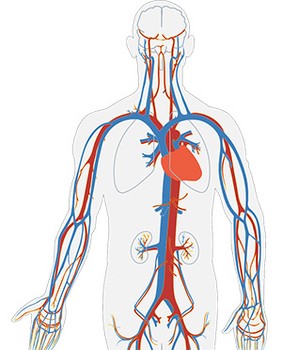
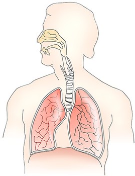
Figure 2. Visualizations of the circulatory system (left) and the respiratory system (right).
- Focus on the respiratory system in the next part of the lesson. Tell students that in this lesson, they will look into the respiratory system in more detail.
How would you define the respiratory system? What is its function and can you list some parts of the respiratory system?The respiratory system is the system responsible for breathing. It consists of the lungs, and the nose, the bronchi, the trachea or windpipe, etc. Slide 3 of the Slideshow can help students visualize these parts. Together with students, define the respiratory system as the collection of parts that are responsible for breathing, or for taking in oxygen and releasing carbon dioxide.
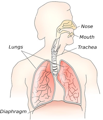
Figure 3. Visualizations of the major parts of the respiratory system.
/-/https/i.ytimg.com/vi/E0yxTOkhzSI/maxresdefault.jpg)
/-/https/www.sciencebuddies.org/Files/14174/23/note-question-mark-30.png)
/-/https/www.sciencebuddies.org/Files/14175/24/conversation-bubbles-30.png)
/-/https/www.sciencebuddies.org/Files/17093/6/visual-circulatory-system.jpg)
/-/https/www.sciencebuddies.org/Files/17094/6/respiratory-system-unlabeled.jpg)
/-/https/www.sciencebuddies.org/Files/16839/7/anatomy-respiratory-system.jpg)
Explore (45 minutes)
- Explain to students that they will be doing an activity to learn more about
the different parts of the respiratory system, their functions, and how they work together.
Introduce the activity:
Show the model on Slide 4 of the Slideshow or the real example you have, and let student groups discuss how it is similar to the respiratory system and write their ideas on their Student Worksheet.
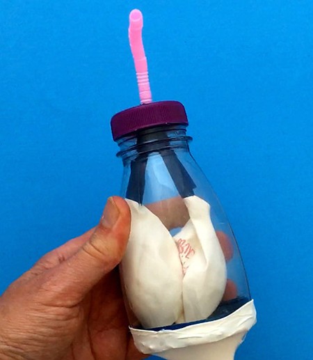
Figure 4. A model of the human respiratory system.Distribute materials while the students discuss in their groups.
- Guide groups through the procedure of building their model.
Show students the video of how to build the model step-by-step. Walk around and assist where needed. If students struggle to make the trachea split into the bronchi, allow them to make a model with one lung, but still ask them to include the trachea.
-
Let groups use the model to discover how the breathing mechanism works.
Let groups make a hypothesis of how breathing (air flowing in and out of the lungs) in humans works using their model to support their hypothesis (worksheet question 4).
Discuss some of the hypotheses as a class.
End this step by explaining that when we breathe in, air automatically flows into our lungs because the diaphragm flattens. That is like us pulling the knot back in the model. When we pull the knot back, air flows into the balloons. To breathe out, the diaphragm relaxes again, pushing air out of the lungs. In our model, we release the knot and gently push it in. We see that air flows out and the balloons empty and start to cave in.
- Help students
discover the specific function and characteristics of different parts of the respiratory system.
- Let groups map parts of the model to parts of the respiratory system and list the function of each part (question 5 on the worksheet).
Swipe left to see more
Table 1. Comparison between the model and the respiratory system it models.Model Function Respiratory system Nose (and mouth) End of the straw Opening that allows air to flow in and out. Trachea (or windpipe) Drinking straw Channels airflow toward and from the lungs to the nose (and mouth). Bronchi Branched-off drinking straws Channel the airflow toward or from one of the two lungs. Lungs Balloons Allow exchange of oxygen and carbon dioxide with blood. Diaphragm Balloon membrane with knot Allows an increase and decrease in the volume of the lungs so air flows in and out. Other systems interacting with the respiratory system Ribcage Hard plastic bottle Protects the lungs and heart. Blood Not modeled Transports oxygen from the lungs to the cells, and carbon dioxide from the cells to the lungs.
- Let groups think about the shapes and material used for two parts in the model: are they well-chosen; do they support the function of the part?
- Let groups think about these two parts in the human body and wonder if and why these parts are made of the same tissues. Students can find the definition of "tissue" as "a group of similar cells that are specialized to perform a specific function. " as well as the definition of "cell" on their worksheet.
- End this step by showing the students how one could have mapped the parts of the model to the parts of the respiratory system (see Slide 5 of the Slideshow) and ask if students came up with other ways to map it. Listen and discuss their choices, if they had other choices, and go over the function of each part. An example of a complete table is included as Slide 6.
Which groups thought that their two chosen body parts would be made up of the same tissues?Listen to these groups' parts and the reasoning behind their conclusion. As you listen, make sure they understand the term "tissue" as a group of similar cells that are specialized to perform a specific function.What groups thought that their two chosen body parts would be made up of different tissues?Listen to these groups' answers. If there is a lot of controversy, consider looking up the tissues of different parts of the respiratory system; for example, the lungs are made up of lung tissue, the diaphragm is skeletal muscle tissue together with a tendon that anchors the diaphragm to the ribs, the bronchi are mainly made up of cartilage and smooth muscle tissue, the larynx is mainly made of cartilage, etc.
/-/https/www.sciencebuddies.org/Files/17095/6/lung-model-example.jpg)
/-/https/i.ytimg.com/vi/mfiWzupeK3I/maxresdefault.jpg)
/-/https/www.sciencebuddies.org/Files/17096/6/muscle-tissues.jpg)
/-/https/www.sciencebuddies.org/Files/17097/10/lung-tissue-horiz-2.jpg)
Figure 5. Left: Picture of the three types of muscle tissue found in the human body: (a) skeletal muscle tissue, (b) smooth muscle tissue, and (c) cardiac muscle tissue. (Same magnification.) Right: Picture of lung tissue. (500X magnification).
- Tell students that all of these tissues consist of cells. The cells from different tissues can look different and can have a different function, but they are all living cells. Ask students to identify some cells in Slide 7 of the Slideshow (Figure 5). The dark spots are the nuclei of the cells. This slide shows muscle and lung tissue under a microscope and Slide 8 of the Slideshow (Figure 6) shows what a cell might look like. Conclude that the respiratory system consists of different parts and different tissues. Each part or each type of tissue is specialized in a specific function, but they all work together to perform one task.
/-/https/www.sciencebuddies.org/Files/17098/6/cell-graphical.jpg)
Figure 6. A graphical representation of a cell.
- Discover how the respiratory system works together with the circulatory system.
We have seen that the function of the respiratory system is to absorb oxygen and release carbon dioxide. Can you come up with a hypothesis of how the respiratory system works together with at least one other body system to fulfill its function?Let students discuss this question in their groups and write their hypothesis on the worksheet before addressing this question as a class. Listen to students' answers. Make sure to include the circulatory system, as it helps distribute the oxygen all over the body. Carbon dioxide is transported in a similar way back to the lungs. Students might also mention the skeletal system, as the ribcage is there to protect the lungs. It also holds people upright, creating the space for the chest cavity. The muscular system helps expand and contract the lungs more fully, and the nervous system transmits the signals that initiate breathing; even involuntary breathing needs signals.Do you know how red blood cells obtain and transport the oxygen we breathe in?Listen to students' answers and fill in where needed. In our model, the lungs are big bags, but real lungs are full of tiny sacs called alveoli. The walls of the alveoli have a lot of very fine blood vessels called pulmonary capillaries, as can be seen in Figure 7 (slide 9 of the Slideshow). Oxygen can pass through the thin walls and dissolve in the blood within the capillaries. Some of the oxygen then binds to hemoglobin, a protein that is inside red blood cells. There are about 270 million hemoglobin proteins inside one red blood cell, and there are about 5 million red blood cells in 1 mL of blood (see Figure 8, slide 10 of the Slideshow). The more hemoglobin you have, the easier it is for your body to transport oxygen.
/-/https/www.sciencebuddies.org/Files/17099/6/lung-structure-normal.jpg)
Figure 7. Detailed view of the lungs.
/-/https/www.sciencebuddies.org/Files/17100/6/blood-in-artery.jpg)
Figure 8. A representation of red blood cells.
Reflect (15 minutes)
- Summarize what was learned.
Use the following questions to recapitulate what was learned.
We started with the respiratory system and broke it down into smaller and smaller parts. What was the smallest living unit in the human body we discussed? What was the next level up, and the next level, etcetera, all the way to the human body?The smallest living unit was the cell. Some students might even go smaller and mention the proteins that can be carried in the cell (e.g., the hemoglobin carried in the red blood cells). Confirm that these proteins are smaller and add that the cell is the smallest living unit. Protein are molecules, they are not living. We then grouped similar cells that are specialized in a specific function into tissues, and tissues are sometimes part of an organ. We grouped body parts and organs that together fulfill an important function for the organism into systems, and systems work together to create the organism. - Ask students to order the following words from least-complex to most-complex on the worksheet before addressing the answer as a class.
- Lung, lung cell, human, lung tissue, respiratory system
- Tissue, system, organ, cell, organism
- Come back to question 1 on the Student Worksheet where you asked students about the connection between iron deficiency anemia and shortness of breath.
Looking at everything you learned in this lesson, and knowing that you need iron to create hemoglobin, would you change your answer to question 1?Let students write down their answer on the worksheet, or give this question as homework, before addressing it as a class. Guide students to conclude that if you do not have enough iron, you are not able to make sufficient hemoglobin and because of it, the red blood cells pick up less oxygen. Since there is less oxygen in the blood, the cells in the body do not as easily get the oxygen they need to work efficiently. Your body makes adjustments as it tries to get enough oxygen to the cells. For example, your heart might start pumping faster in an attempt to circulate blood faster, and thus circulate oxygen faster. You might also start to breathe faster, or feel out of breath, as your body tries to inhale more oxygen.
- End the lesson by asking students to write down something they learned, something that surprised them, and something they would like to know more about.
Assess
Questions 10 and 11 of the worksheet can be used to assess the students' understanding.
You can use this quiz to assess student learning after the activity:
- Online quiz, assignable in any LMS
- Quiz (pdf) and answer key (PDF)
Make Career Connections
Discussing or reading about these careers can help students make important connections between the in-class lesson and STEM job opportunities in the real world.
/-/https/careerdiscovery.sciencebuddies.org/Files/979/18/pexels-photo-3912962.jpg)

/-/https/careerdiscovery.sciencebuddies.org/Files/967/17/unsplash-d4KQc-ZqBrc.jpg)
Lesson Plan Variations
- Instead of the respiratory system, examine a more generally known system outside of the human body and decompose it into its less-complex parts. Rails are basic building blocks of tracks that can belong to a railway system; a brick is the basic building block of a wall, which can be assembled into a room; and a collection of rooms can serve as a house. Each part fulfills a specific task in the whole. Compare these organizations with the human body.
- Choose to model a different body system or have different groups in the class choose a different body system to explore.
- Look at the animal kingdom. Can you find a similar organization of cells into tissue, organs, and systems there?
- Instead of this more-complicated lung model, build the simple lung model, as explained in the How Do We Breathe? activity. Examine what parts have been left out and wonder what the advantage could be of having these parts.
/-/https/img.youtube.com/vi/JrV_f_omYHY/0.jpg)
/-/https/img.youtube.com/vi/TsgryTenm04/0.jpg)
/-/https/img.youtube.com/vi/u3LdI00902o/0.jpg)